RESEARCH
During my first February internship at the NYU Barrett Lab under supervisor Dr. Tessa Barrett and mentor Dr. Min Dai, I analyzed a variety of protein datasets. I processed data, exported measurements, and built figures in GraphPad Prism. Creating publication-style graphs was genuinely fun, especially experimenting with axes to view the data from new perspectives that could highlight otherwise overlooked discrepancies. It was also satisfying knowing the figures could be presented at lab meetings to communicate results and support statistical conclusions.
I was also responsible for helping transition the lab’s data into Benchling. I documented protocols and materials so they could be easy to access in the new system, and I updated them as the project evolved, for example adjusting reagent amounts as conditions became more defined. This work required careful chemistry calculations to scale materials accurately. The lab team used my calculations to prepare the amount of compound needed to generate enough cells to freeze for later data collection.
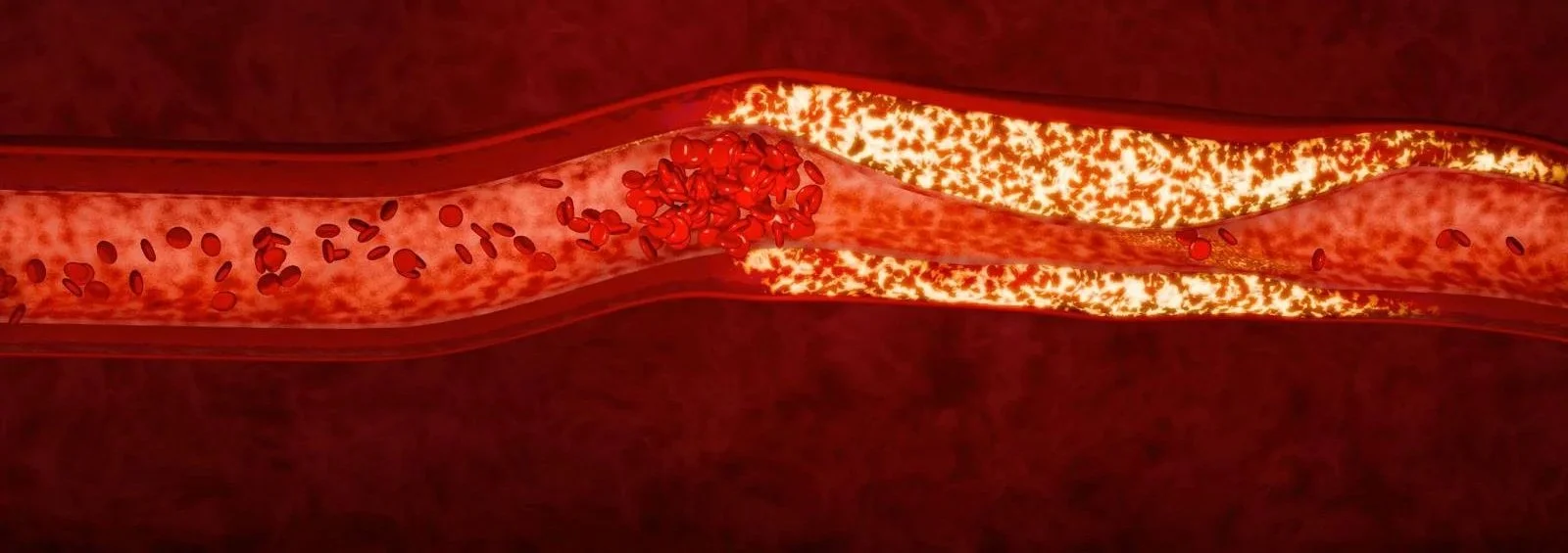
This past summer, I worked full-time as a researcher alongside two undergraduate researchers studying platelet activity in atherosclerosis. Seeing the full research arc, from confirming that samples were ready for imaging, to image acquisition, to analysis, helped me understand how each step supports the larger picture. Our project examined how the protein CD37 is involved in plaque buildup after damage to the aorta. Platelets are best known for stopping bleeding, but they also play key roles in inflammation, immunity, and tissue repair. Our goal was to understand how platelet signaling contributes to cardiovascular disease so we can eventually develop safer, more targeted treatments for conditions like heart attacks, stroke, and atherosclerosis.
My responsibilities included collecting images, cutting out pieces of plaque, and quantifying plaque buildup in each aorta. I appreciated learning techniques that made our data strong from the start, and operating the imaging system was especially meaningful. Given a prepared sample on a plate, I loaded it into the instrument, located the regions we needed to capture, and acquired both nonfluorescent and fluorescent images using the control panel. I verified resolution and focus and avoided artifacts such as air bubbles. Completing careful image acquisition taught me how reliable inputs are the foundation for everything that follows. On the analysis side, I used Fiji/ImageJ to process images and quantify signals, including estimating the percent area positive for CD37 in atherosclerotic plaque sections in a stress model of mice. This contributed to exploring whether stress is associated with increased CD37 signal in plaques.
It was enriching to work alongside graduate-level members and staff and to contribute reliably in a collaborative, fast-paced environment, and I’m confident that is where I want to be working in the future. My contribution this summer, a repeatable workflow for quantifying aortic plaque, earned me an invitation to return for future bioinformatics projects. To prepare, I am currently conducting independent work on bioinformatics pipelines that I will apply during my summer 2026 research.